Введение
Совмещенная позитронно-эмиссионная и компьютерная томография (ПЭТ/КТ) – наукоемкий высокотехнологичный метод структурно-функциональной лучевой визуализации физиологических и биохимических процессов в организме здорового и больного человека. Она применяется для раннего выявления опухолевых образований, диагностики жизнеспособности миокарда, воспалительных заболеваний сердца и сосудов, психоневрологических расстройств и т.д. С помощью этого метода можно количественно оценивать состояние метаболизма на клеточно-молекулярном уровне и фиксировать тонкие биохимические изменения определяющих основу различных патологически состояний [1, 2].
Основу метода составляет специальная маркировка биомолекул радиофармацевтическими препаратами (РФП) для визуального выявления их гиперметаболизма в опухолевых образованиях или очагах активного воспаления, а также гипо- или изометаболизма в зонах диффузного хронического поражения тканей с одновременным математическим расчетом цифровых значений показателя захвата РФП (SUVmax) в условных единицах при общепринятых значениях 2,0≥ у.е. [3, 4].
Согласно существующим рекомендациям по ПЭТ/КТ-обследованию онкологических пациентов, высокая метаболическая активность в отношении 18F-ФДГ-глюкозы характерна для опухолевых клеток меланомы кожной, а 11С-холина – для раковых клеток предстательной железы. Чувствительность метода составляет 96%, диагностическая точность – 97,2% [5, 6].
В то же время структурно и функционально состоятельные центры головного мозга, координирующие работу нижних мочевых путей в фазу наполнения и опорожнения мочевого пузыря, также на локальном уровне проявляют высокую метаболическую активность в отношении глюкозы, что позволяет визуализировать и математически рассчитывать интенсивность этого процесса [7–9].
 Не значит ли это, что все органоспецифические клетки органов мочеобразования и выведения мочи в процессе обследования на наличие онкологических заболеваний также могут принимать участие в метаболизме используемых РФП, исходя из своих физиологических потребностей?
Цель исследования: изучить возможность анализа структурно-функционального состояния почек и мочевыводящих путей по результатам ПЭТ/КТ-исследования всего тела у пациентов с меланомой кожи и раком предстательной железы в стадии ТIN0M0 без признаков метастазирования и наличия нефроурологического анамнеза.
Материал и методы
Проведен ретроспективный анализ 50 ПЭК/КТ-заключений больных указанных групп, выполненных за 2016–2018 гг. в Радиологическом центре Тюменского областного онкологического диспансера по стандартной методике на аппарате PET/CT (Siemens Biograph) производства Германии. Отбирались заключения пациентов среднего возраста с медианой 39,5±1,4 года, с отсутствием ПЭТ/КТ-признаков метастазирования и нефроурологического анамнеза. В 25 случаях исследование проводилось с 18ФДГ-глюкозой и в 25 случаях – с 11С-холином. Зоны интереса анализировались полуколичественным методом и картировались штрих-линией. В этих зонах вычисляли значение уровня захвата изотопа (SUVmax) в условных единицах. Расчет проводился программным комплексом автоматически. Материалы исследования были обработаны и систематизированы с применением программного пакета электронных таблиц Microsoft Exel и пакета программ «Statistica 10».
Результаты исследования
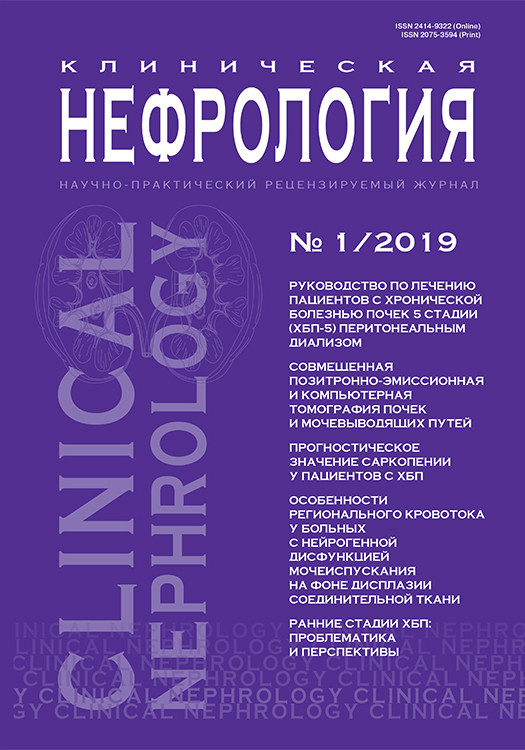
На рис. 1. представлены КТ- и ПЭТ/КТ-томограммы почек и мочевыводящих путей пациентов с применением различных РФП. Представленные данные позволяют сделать вывод, согласно которому ПЭТ/КТ-визуализация структурно-функционального состояния почек и мочевыводящих путей возможна и при этом достаточно информативна.
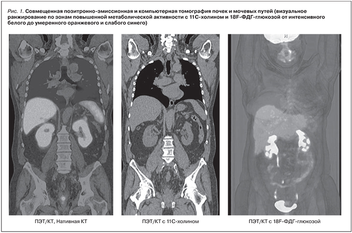
На рис. 2. представлены варианты ПЭТ/КТ-томограмм пациентов с расчетом показателя захвата изотопа в корковом, мозговом слоях паренхимы, а также в полости лоханок структурно и функционально состоятельных почек.
Математический расчет уровня захвата РФП в корковом, мозговом слоях почек, а также проекции лоханок позволил оценить у этих пациентов структурно-функциональное состояние почек в процессе мочеобразования и выведения мочи как удовлетворительное не только визуально, но и в цифровом выражении ( SUVmaх 2,0≥ у.е. ), а также сделать это раздельно для каждого органа.
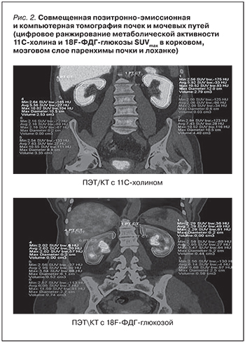
На рис. 3. представлены некоторые варианты структурно-фукциональной нефроурологической патологии, впервые выявленной у пациентов в процессе проведенного ПЭТ/КТ-исследования.
Выводы
Проведенные исследования позволяют сделать вывод, согласно которому не только высокоточная диагностика онкологических заболеваний, оценка состояния жизнеспособности миокарда, а также визуализация и расчет активности функционально состоятельных центров коры головного мозга в процессе реализации накопительно-эвакуационных функций нижних мочевых путей, но и структурно-функциональные изменения в паренхиме почек, верхних мочевых путях в процессе образования и выведения мочи могут быть визуально и математически проанализированы методом совмещенной ПЭТ/КТ.